
Education
12 Lead ECG Placement and how to use ECG device
An electrocardiogram, or ECG, is a reading assessing the magnitude and direction of the electrical currents of the heart, and measuring the depolarisation and repolarisation of the cardiac muscle cells (Medani et al 2018).
It is important an ECG is recorded accurately.
ECG electrode placement is standardised, allowing for the recording of an accurate trace – but also ensuring comparability between records taken at different times.
Poor electrode placement can result in mistaken interpretation, and then possible misdiagnosis, patient mismanagement and inappropriate procedures (Khunti 2013). Deviation of lead placement even by 20-25mm from the correct position can create clinically significant changes on the ECG, including changes to the ST-segment (McCann et al. 2007).
Patient factors may also contribute to the variability in accuracy, such as the patient’s respiration, position, smoking, recent dietary intake and obesity (McCann et al. 2007).
It is therefore important to not only ensure that the electrodes are placed in accordance with the standardised ‘rules’ but also, that the patient is prepared correctly, both physically and psychologically for the procedure.
Preparing a Patient for an ECG
As with all procedures, we should obtain informed consent from the patient; explaining the purpose of the procedure; the procedure itself and obtaining consent to proceed. Maintain good infection control practice by washing your hands prior to patient contact.
Skin preparation is important. If the patient’s skin is dirty, clean with soap and water, and then dry. If the skin is oily or the patient applied any creams or lotions, use an alcohol wipe to clean each electrode placement site.
Some ECG machines may also provide a ‘rough patch’ either separately or on the electrodes, which can be used to rub on the skin to increase electrode adherence, but care should be taken not to cause abrasions.
Patients with chest hair should have hair at the electrode placement sites removed with a razor (Coviello 2016).
Where possible, place the patient in a semi-recumbent position (Baillie 2014). If this is not possible or uncomfortable for the patient, it is acceptable to record the ECG in another position.
The patient must be completely relaxed. Ensure the environment is at a comfortably warm temperature (Jevon 2010). This will prevent muscular tension or movements producing artefact on the ECG recording. Ensure privacy and dignity: e.g. closing
the room door or drawing around the curtains.
12-Lead ECG Placement
The patient’s chest and all four limbs should be exposed in order to apply the ECG electrodes correctly.
There are different methods for identifying the correct landmarks for ECG electrode placement, the two most common being the ‘Angle of Louis’ Method and the ‘Clavicular’ Method (Crawford & Doherty 2010a).
Regardless of the method used, the ECG electrode positions should be found in the following locations:
| V1 (C1) | Fourth intercostal space at the right sternal border |
|---|---|
| V2 (C2) | Fourth intercostal space at the left sternal border |
| V3 (C3) | Halfway between leads V2 and V4 |
| V4 (C4) | Fifth intercostal space in the midclavicular line |
| V5 (C5) | Left anterior axillary line on the same horizontal plane as V4 |
| V6 (C6) | Left midaxillary line on the same horizontal plane as V4 and V5 |
| RA (R) | Right arm (inner wrist) |
| LA (L) | Left arm (inner wrist) |
| RL (N) | Right leg (inner ankle) |
| LL (F) | Left leg (inner ankle) |
| Unbracketed letters indicate lead names under the American Heart Association (AHA) system; bracketed letters indicate lead numbers under the International Electrotechnical Commission (IEC) |
Precordial Lead Placement
In order to find these correctly, the ‘Angle of Louis’ Method can be used:
- To locate the space for V1; locate the sternal notch (Angle of Louis) at the second rib and feel down the sternal border until the fourth intercostal space is found. V1 is placed to the right of the sternal border, and V2 is placed at the left of the sternal border.
- Next, V4 should be placed before V3. V4 should be placed in the fifth intercostal space in the midclavicular line (as if drawing a line downwards from the centre of the patient’s clavicle).
- V3 is placed directly between V2 and V4.
- V5 is placed directly between V4 and V6.
- V6 is placed over the fifth intercostal space at the mid-axillary line (as if drawing a line down from the armpit).
- V4-V6 should line up horizontally along the fifth intercostal space.(Coviello 2016)
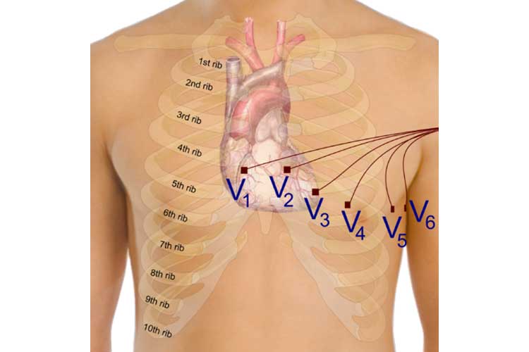
The ‘Angle of Louis’ Method can be used in the placement of the precordial electrodes.
The Clavicular Method is useful as a check, but Crawford and Doherty (2010b) advise that inexperienced practitioners may mistake the sub-clavicular space as the 1st intercostal space; meaning that the intercostal spaces are incorrectly identified.
Limb Lead Placement Diagram
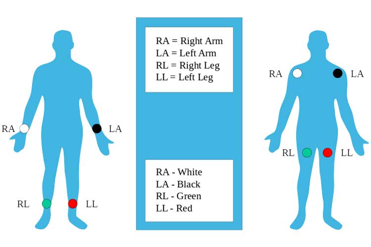
The limb electrodes can be far down on the limbs or close to the hips/shoulders as long as they are placed symmetrically.
Other Considerations
Breast tissue can impact on the ECG amplitude due to the increased distance between the electrode and the heart when ECG electrodes are placed over the chest (Rautaharuju et al. 1998).
The V4 lead is recommended to be placed underneath the breast tissue in women. Crawford and Doherty (2010b) point out that it makes little sense to locate the correct position under the breast, to then replace the breast and attempt to approximate the correct location. Thus, they recommend that V4 should be placed under the breast, and V5 and V6 placed underneath too if lifting the breast is needed.
It is often customary in practice to write on the ECG if an electrode has been placed over breast tissue in order to aid the interpretation.
Where it becomes necessary, it is also customary practice to record any alterations in lead placement; for example, where lead placement is changed from the standardised location due to patient position, injury etc.
End of Procedure
Ensure that the patient’s privacy and dignity is maintained. The chest should not be left exposed and can be covered back up with blankets, or allow the patient to re-dress as necessary.
The ECG electrodes should be removed if the patient is not likely to require further or serial ECGs, but otherwise can be left in place for up to 24 hours before needing to be replaced (Coviello 2016).
If you are not interpreting the ECG, follow local policy and use clinical judgement to arrange for interpretation. Local policies often also require the initials of the person taking the ECG to be recorded.
References:
- Baille, L 2014, Developing practical nursing skills, 4th edn, CRC Press, Florida.
- Coviello, J 2016, ECG Interpretation made incredibly easy, 6th edn, Wolters Kluwer, London.
- Crawford, J & Doherty, L 2010a, ‘Ten steps to recording a standard 12-lead ECG’, Practice Nursing, vol. 21, no. 12, pp. 622-29, viewed 13 March 2018, https://www.magonlinelibrary.com/doi/abs/10.12968/pnur.2010.21.12.622.
- Crawford, J & Doherty, L 2010b, ‘Recording a standard 12-lead ECG: Filling in gaps in knowledge’, Journal of Paramedic Practice, vol. 2, no. 3, pp. 102-8, viewed 13 March 2018, https://www.magonlinelibrary.com/doi/10.12968/jpar.2010.2.3.47285
- Jevon, P 2010, ‘Procedure for recording a standard 12-Lead electrocardiogram’, British Journal of Nursing, vol. 19, no. 10, pp. 649-51, viewed 13 March 2018, https://www.ncbi.nlm.nih.gov/pubmed/20622761
- Khunti, K 2013, ‘Accurate interpretation of the 12-lead ECG electrode placement: A systemic review’, Health Education Journal, vol. 73, no. 5, pp. 610-23.
- McCann, K, Holdgate, A, Mahammad, R & Waddington, A 2007, ‘Accuracy of ECG electrode placement by emergency department clinicians’, Emergency Medicine Australasia, vol. 19, pp. 442-8, viewed 13 March 2018, https://www.ncbi.nlm.nih.gov/pubmed/17919217
- Medani, S, Hensey, M, Caples, N & Owens, P, 2018, ‘Accuracy in precordial ECG lead placement: improving performance through a peer-led education intervention’, Journal of Electrocardiology, vol. 51, pp. 50-4, viewed 13 March 2018, https://www.ncbi.nlm.nih.gov/pubmed/28576322
- Rautaharju, P, Park, L, Rautaharju, F & Crow, R 1998, ‘A standardized procedure for location and documenting ECG chest electrode positions: Consideration of the effect of breast tissue on ECG amplitudes in women’, Journal of Electrocardiology, vol. 31, no. 1, pp. 17-29, viewed 13 March 2018, https://www.ncbi.nlm.nih.gov/pubmed/9533374.
Promoted Products
Meditech Brands


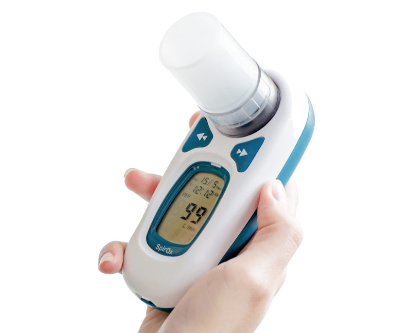
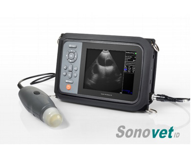
Meditech Equipment Co.,Ltd is part of Meditech Group. Product(s) described may not be licensed or available for sale in all countries. Sonotech, Sonovet, iSonic, FOs2pro, Dolphi, Defi, HeartRec,miniScan,Cardios,SpirOx,iBreath, Meditech and all corresponding design marks are trademarks of Meditech. The symbol indicates the trademark is registered. Patent and Trademark Office and certain other countries. All other names and marks mentioned are the trade names, trademarks or service marks of their respective owners. Please see the Instructions for Use for a complete listing of the indications, contraindications, warnings and precautions.
Legal notice Terms and conditions Cookie policy Privacy Policy Professional organisations Careers