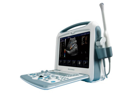
Probes Made by NDK JAPAN
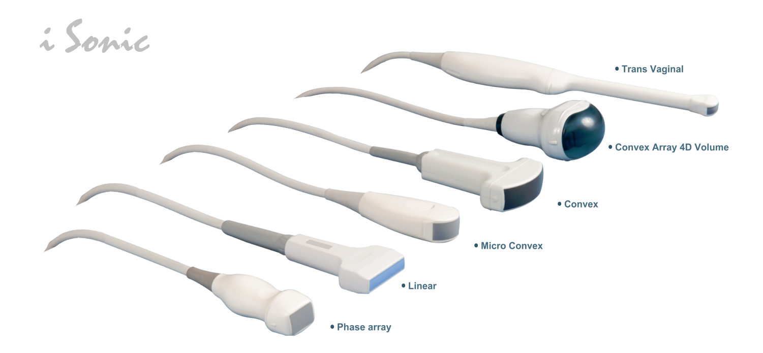
Product Features
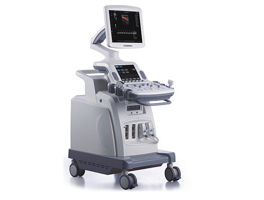
iSonic® Wide band multi frequency ,Imaging Processing ,Imaging optimization
195 elements, ZIP260 connector
Hard Disk (500G),
Control panel (exclude touch pad)
Operating Mode: 2B, 4B, B/M, M, B/C, B/C/D, B/D, PW, velocity,
power (direction), histogram, Triplex/Duplex
|
Imaging Processing Technology Imaging optimization technology Compound enhance technology Speckle reduction Multi beam parallel processing technology Color coding Doppler frame correlation Wall filter Tissue Harmonic |
File management Hard disk storage Cine loop DVD-ROM USB RS232 DICOM 3.0 Intranet Parallel printing port |
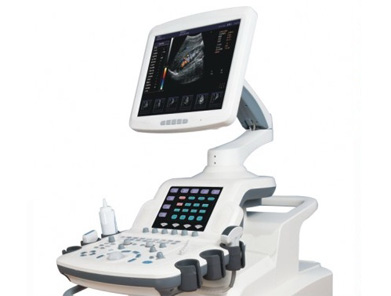
Imaging Modes
Overview
B, Dual B, Quad B, M
Color, Dual Color
Simultaneous 2D/Color Compound
PW, Duplex/Triplex
CFM, PD, Directional PD, CD
Freehand 3D
4D
Panoramic
Measurements Specifications
Obstetrical Report
Amniotic Fluid Index (AFI)
BPD/OFD, FL/AC, FL/BPD and HC/AC ratio
Estimate Weight Of Fetus
Gestational Age
Expected Date Of Confinement (LMP/BBT)
Fetal Biophysical Score
Fetus Growth Curve
Gynecology
Uterus, Ovary, Follicle
Gynecology Report
Urology
Kidney, Bladder, Residual Urine Volume
Urology Report
Andrology
Prostate, Testis
Prostate Specific Antigen (PSA)
Prostate Specific Antigen Density (PSAD)
Andrology report
Orthopedic
Left and Right Hip Joint
Peripheral Vascular
Area Stenosis
Vessel Diameter Stenosis
Peripheral Vascular Report
Small Parts
Thyroid, Mammary Gland, Nodule
Small Parts Report
Multiple Births
Measurement Of Multiple Births
Cardiac
Heart Rate
Blood Flow Rate
Left Ventricle
Aorta
Mitral Valve
Ventricle (left/right)
Area Stenosis (% area Sten)
Vascular Diameter Stenosis (% Diam Sten)
Body Surface Area (BSA)
Product Specifications
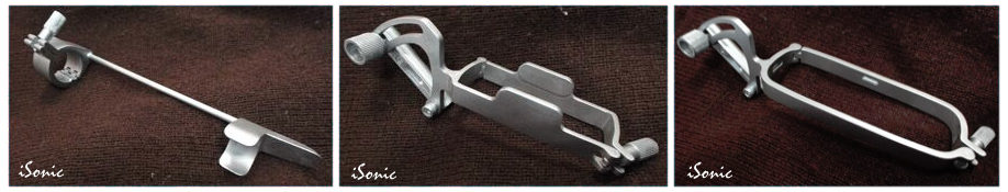
Related Accessories
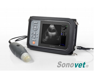
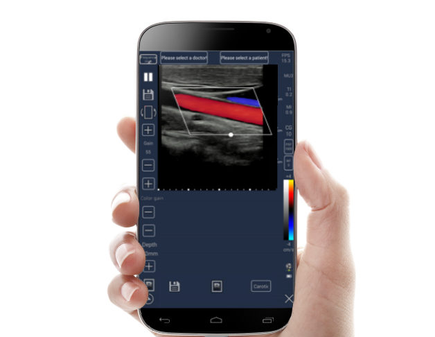
Related Products
Promoted Products
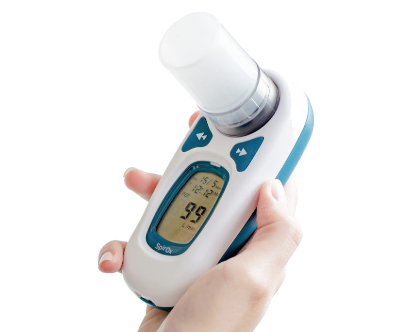
Spirometer Type: P
Palm size Spirometer with LCD screen PEF and FEV1 test Meet ATS American Thoracic Society ,1994 update standardization. Suitable for hospital or home. Maximum of records is 600 measurements
Meditech Equipment Co.,Ltd is part of Meditech Group. Product(s) described may not be licensed or available for sale in all countries. Sonotech, Sonovet, iSonic, FOs2pro, Dolphi, Defi, HeartRec,miniScan,Cardios,SpirOx,iBreath, Meditech and all corresponding design marks are trademarks of Meditech. The symbol indicates the trademark is registered. Patent and Trademark Office and certain other countries. All other names and marks mentioned are the trade names, trademarks or service marks of their respective owners. Please see the Instructions for Use for a complete listing of the indications, contraindications, warnings and precautions.
Legal notice Terms and conditions Cookie policy Privacy Policy Professional organisations Careers